Many individuals suffering from obstructive sleep apnea often face the dilemma of choosing the most suitable Continuous Positive Airway Pressure (CPAP) mask for their treatment. Two popular options are full-face and nasal CPAP masks. This article delves into the differences between these two types, aimed at helping you make an informed decision.
Understanding CPAP masks and their purpose
CPAP therapy is widely regarded as a highly effective treatment for sleep apnea, a condition characterised by interrupted breathing during sleep. The primary purpose of a CPAP mask is to deliver a continuous stream of air to help keep the airway open, thus preventing apnoeic episodes.
The role of CPAP masks in sleep apnea treatment
Utilising a CPAP mask is essential for those diagnosed with sleep apnea. By maintaining a steady airflow, best cpap masks significantly enhance the quality of sleep and reduce health risks associated with untreated apnoea, such as cardiovascular issues and daytime fatigue.
The mask’s design and fit play a pivotal role in the overall effectiveness of the treatment. A mask that is neither too tight nor too loose can ensure that the pressure remains consistent throughout the night, allowing for a restorative sleep experience. Furthermore, patients often report that a well-fitted mask not only improves their comfort but also enhances their adherence to therapy, which is crucial for long-term health benefits.
The basic design and function of CPAP masks
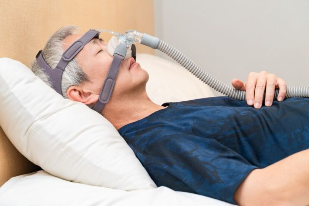
Generally, CPAP masks are crafted from soft, flexible materials designed to contour to the face. They come in various styles, including full-face, nasal, and nasal pillow masks, each designed for specific needs and preferences. The mechanism behind them involves a connection to a CPAP machine that generates air pressure. This consistent pressure prevents the airway from collapsing during sleep, which is the crux of sleep apnea treatment.
Individuals must choose a mask that fits securely yet comfortably to ensure compliance with the therapy. A good fit is essential for effective treatment and to reduce common issues such as skin irritation or discomfort during use. Additionally, many modern masks incorporate features such as adjustable straps and breathable fabrics to enhance comfort further. Some users may also benefit from heated humidifiers that can be attached to their CPAP machines, which help to alleviate dryness in the airways and make the experience more pleasant.
Moreover, the choice of mask can also be influenced by lifestyle factors. For instance, active individuals or those who tend to change sleeping positions frequently may prefer nasal pillow masks due to their lighter weight and less obtrusive design. Conversely, full-face masks may be more suitable for patients who breathe through their mouths or require higher pressure settings. Understanding these nuances can empower users to make informed decisions about their CPAP therapy, ultimately leading to better health outcomes and improved quality of life.
The characteristics of full-face CPAP masks
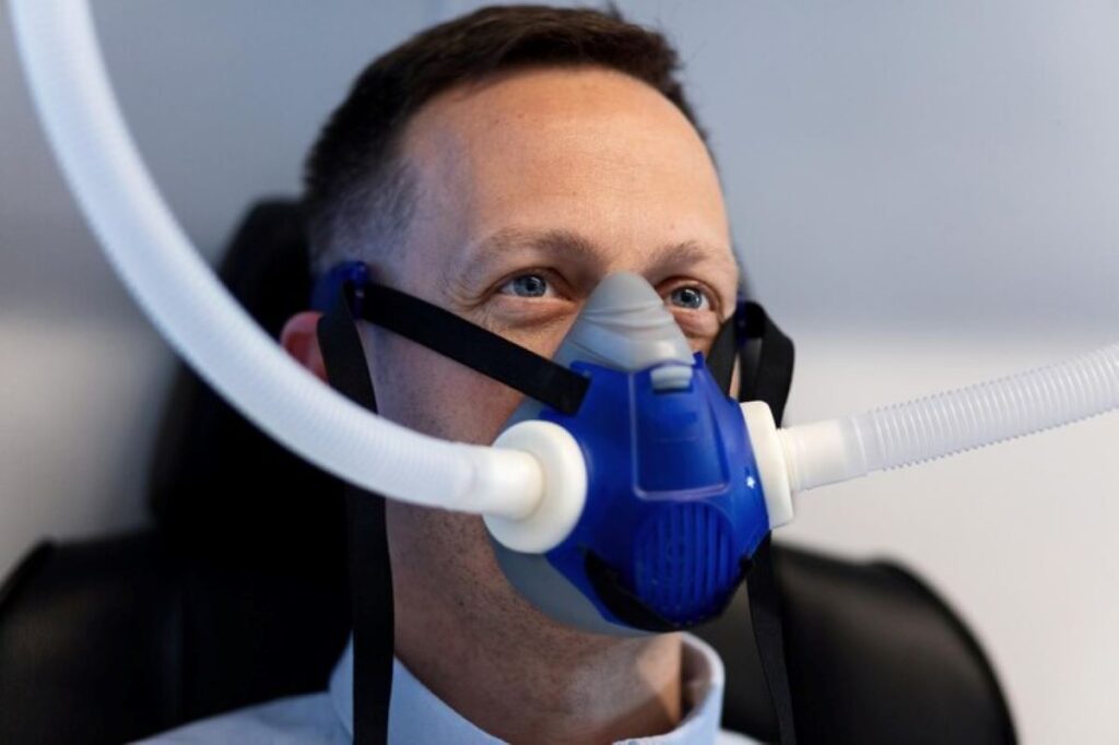
Full-face CPAP masks cover both the nose and mouth, making them an excellent choice for individuals who predominantly breathe through their mouths during sleep. Understanding their design and functionality can help potential users determine if this is the right option for them.
The design and fit of full-face masks
Full-face masks are generally larger compared to their nasal counterparts and require proper sizing for an effective seal. They feature adjustable straps, enabling users to achieve a snug fit without excessive pressure on the facial skin.
Moreover, some full-face masks come with a cushion that creates a seal around the face, minimising air leaks. This ensures that users receive optimal pressure levels throughout the night. The materials used in these masks are often soft and hypoallergenic, which can be particularly beneficial for those with sensitive skin. Additionally, many modern designs incorporate a lightweight frame that reduces the overall bulk, making them more manageable for wearers during sleep.
The benefits and drawbacks of full-face masks
One of the significant benefits of full-face masks is their versatility. Those who suffer from nasal congestion or have a tendency to breathe through their mouth can benefit greatly from this design. Moreover, full-face masks generally provide a more stable airflow, reducing the risk of apnoea episodes during the night. Users often report a more consistent and uninterrupted sleep experience, as these masks are less likely to dislodge during movement, ensuring that the therapy remains effective throughout the night.
However, there are some drawbacks as well. Users may find full-face masks to be bulkier and less comfortable than nasal options. They can also lead to greater feelings of claustrophobia for some individuals, which may interfere with achieving restful sleep. Additionally, the larger surface area can sometimes cause skin irritation or pressure sores if not fitted correctly. Regular cleaning and maintenance are essential to prevent the build-up of bacteria and ensure the longevity of the mask, as neglecting these aspects can lead to discomfort and health issues over time.
The specifics of nasal CPAP masks
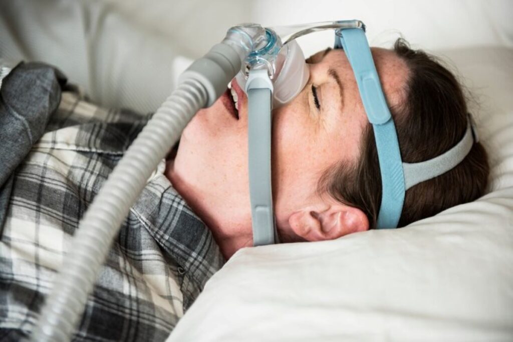
Nasal CPAP masks, on the other hand, cover only the nose and are popular among those who prefer a lighter, less intrusive option. Understanding their design and utility is essential for making a well-informed decision.
The design and fit of nasal masks
Nasal masks are designed to distribute pressure evenly across the nose, which makes them generally lighter and less cumbersome. They often feature a minimalist design with fewer components compared to full-face masks. This can translate to easier maintenance and cleaning.
Finding the right size and fit is crucial for nasal masks as well. A snug fit will prevent air leaks and ensure effective treatment, while an overly tight fit can cause discomfort and irritation. Many manufacturers offer a range of sizes and adjustable straps, allowing users to customise their masks for optimal comfort. Additionally, some nasal masks come equipped with memory foam cushions that adapt to the contours of the face, providing a personalised fit that can enhance both comfort and effectiveness.
The advantages and disadvantages of nasal masks
Nasal masks have several advantages. They are typically lighter and less obtrusive, which many users find preferable for sleeping. The smaller profile can also be easier to tolerate for those who feel claustrophobic with larger masks. Furthermore, the design of nasal masks often allows for greater freedom of movement during sleep, enabling users to shift positions without the mask shifting or causing discomfort.
However, such masks do come with drawbacks. Users who tend to breathe through their mouths or have chronic nasal congestion may struggle with maintaining the necessary airflow, thus leading to potential interruptions in therapy effectiveness. To mitigate these issues, some users may opt to use a chin strap to help keep their mouths closed during the night, or they might explore alternative therapies such as humidifiers to alleviate nasal congestion. It’s also worth noting that while nasal masks are generally well-received, individual experiences can vary significantly, making it essential for users to consult with healthcare professionals to find the most suitable option for their specific needs.
Making the choice: Full-face vs nasal CPAP masks
Choosing the right CPAP mask is crucial for your treatment success. Both full-face and nasal masks have their unique characteristics, and individual preferences play a significant role in the decision-making process.
Factors to consider when choosing a mask
Several factors should influence your choice of CPAP mask, including your breathing patterns during sleep, any underlying nasal issues, and your level of comfort. If you often wake up with a dry mouth or experience nasal congestion, a full-face mask may be more beneficial.
Conversely, if you do not experience these issues, a nasal mask might be a sufficient and more comfortable option. Your healthcare provider can provide valuable guidance based on your specific situation. Get about how to get the best fit when you buy CPAP Mask Online on https://ighfamilyeyeclinic.com/how-to-get-the-best-fit-when-you-buy-cpap-mask-online/
The impact of personal comfort and lifestyle on mask choice
Your lifestyle can also dictate the best mask choice. If you are an active sleeper or frequently change positions during sleep, you may require a mask that stays in place effectively. Comfort is paramount; a mask that makes you feel anxious or uncomfortable can lead to non-compliance with therapy.
Moreover, personal habits such as wearing glasses, snoring, or experiencing allergies should also be considered when making your choice. Ensuring that your CPAP therapy fits seamlessly into your life will contribute to better adherence and, ultimately, improved health outcomes.
Frequently asked questions about CPAP masks
It’s common for new CPAP users to have many questions regarding their masks. Addressing these concerns can alleviate apprehension and enhance comfort during treatment.
Addressing common concerns about full-face masks
Many users are often apprehensive about the bulkiness of full-face masks. While it may take some time to adjust, many find that once they acclimatise, they appreciate the stability offered. Regular cleaning and maintenance can also mitigate some common concerns regarding hygiene.
Others may worry about the possibility of skin irritation. Using barrier creams or opting for a mask with soft cushioning can help minimise these issues.
Answering common queries about nasal masks
Common queries about nasal masks often revolve around their fit and potential for leaks. Users are encouraged to try various sizes and styles to find the best match for their facial structure. This is key to achieving the desired therapeutic benefit.
Additionally, though nasal masks are smaller, some users might still experience discomfort or pressure points. Using a nasal mask with adjustable components can alleviate this discomfort and enhance overall satisfaction with CPAP therapy.
In summary, both full-face and nasal CPAP masks have their merits and drawbacks. By understanding the distinctions between them and taking into account individual needs and preferences, users can select the most suitable option. Remember, consultation with a healthcare provider can provide vital insights to help guide your choice.