Deciding to undergo vision correction is a life-changing choice, but for many Australians, the journey starts with a simple, lingering question: Is eye surgery safe? In 2026, the landscape of ophthalmology has reached unprecedented levels of precision, yet outdated fears from the early 2000s still circulate on social media and in dinner party conversations.
Whether you are considering LASIK, SMILE Pro, or advanced lens replacement, understanding the gap between “internet myths” and “medical reality” is essential. This guide breaks down the safety profile of modern eye surgery in the Australian context, providing the clinical clarity you need to move forward with confidence.
See more: SMILE Laser Eye Surgery Reviews: Real Australian Patient Experiences
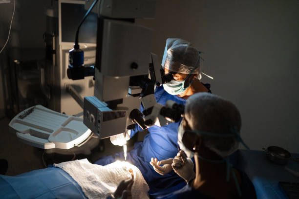
The Evolution of Ocular Safety: Where We Are in 2026
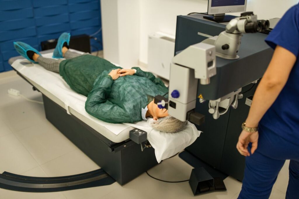
Eye surgery in 2026 is no longer a “one-size-fits-all” laser procedure. It is a highly personalised digital mapping of your unique ocular anatomy. In Australia, clinics now utilise WaveLight ray-tracing technology and AI-driven diagnostic platforms to predict outcomes with microscopic accuracy before a single laser pulse is fired.
The safety of these procedures is anchored by the Royal Australian and New Zealand College of Ophthalmologists (RANZCO) standards, which are among the strictest in the world. Statistically, the risk of a “sight-threatening” complication in a modern Australian clinic is now estimated at less than 0.1%.
5 Common Myths vs. Medical Reality for 2026 Procedures
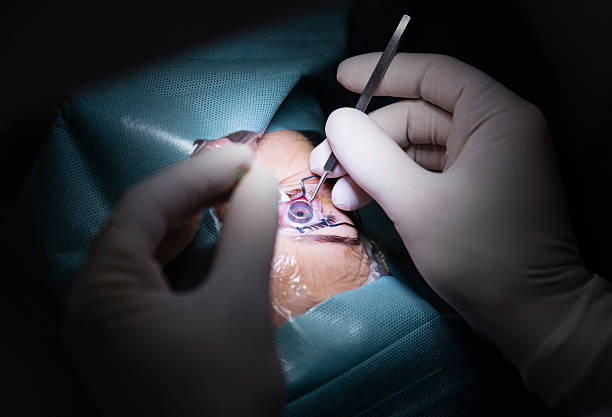
Myth 1: “The laser can burn your eye or face if you move.”
The Medical Reality: Modern lasers, such as the Schwind Atos or VisuMax 800, do not use “heat” in the traditional sense; they are “cold” lasers that break molecular bonds without thermal damage. Furthermore, 2026 technology includes CenTrax® active eye-tracking. If your eye moves even a fraction of a millimetre, the laser compensates instantly or shuts down in a microsecond. It is physically impossible for the laser to “miss” or cause accidental burns.
Myth 2: “You can go blind from laser eye surgery.”
The Medical Reality: There has never been a recorded case of total blindness resulting from a standard LASIK or SMILE procedure in Australia. While all surgery carries a minute risk of infection or corneal ectasia, these are managed through rigorous pre-operative screening. In fact, clinical data suggests that the cumulative risk of a severe eye infection from decades of daily contact lens wear is higher than the one-time risk of modern laser surgery.
Myth 3: “The results don’t last, and you’ll need it again in 5 years.”
The Medical Reality: Laser surgery permanently reshapes the cornea. The physical change to your eye does not “wear off.” However, your eye is a living organ that continues to age. While your distance vision remains corrected, most people will still develop presbyopia (the need for reading glasses) in their mid-40s or cataracts in their 70s. These are natural aging processes, not a failure of the surgery itself.
Myth 4: “The recovery is long, painful, and requires weeks off work.”
The Medical Reality: With the 2026 rollout of SMILE Pro and SmartSight NOVA, the “keyhole” incision is less than 4mm. Most Australians return to office work within 24 to 48 hours. While some mild “grittiness” is common on day one, the intense pain associated with older “surface” procedures (like early PRK) has been largely eliminated by advanced numbing drops and minimally invasive techniques.
Myth 5: “If you have astigmatism or thin corneas, you aren’t a candidate.”
The Medical Reality: This was true 15 years ago. Today, advanced lenticule extraction and Implantable Collamer Lenses (ICL) allow surgeons to treat patients who were previously rejected. High-definition corneal pachymetry (measuring thickness) now identifies the exact safety limits for each patient, making surgery accessible to a much broader range of the Australian population.
Comparison of Leading 2026 Eye Procedures
| Feature | LASIK (Gold Standard) | SMILE Pro (Minimally Invasive) | ICL (Reversible) |
| Best For | Myopia, Hyperopia, Astigmatism | Myopia & Astigmatism | High prescriptions (-10+) |
| Incision Size | ~20mm (Flap) | <4mm (Keyhole) | ~3mm (Lens insertion) |
| Recovery Time | 24 Hours | 24 Hours | 48 Hours |
| Dry Eye Risk | Low | Very Low | Minimal |
| Ideal Candidate | General population | Athletes / Active lifestyles | Thin corneas / High Myopia |
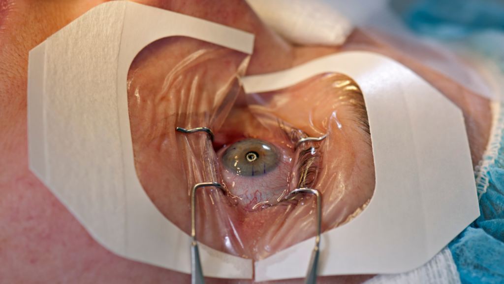
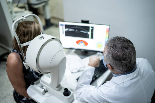
The Step-by-Step Safety Framework in Australia
If you are considering a procedure in Sydney, Melbourne, Brisbane, or Perth, the process follows a strict safety protocol:
- High-Definition Diagnostic Mapping: Using wavefront analysis to create a “fingerprint” of your eye.
- Stability Verification: Confirming your prescription hasn’t changed in the last 12–24 months.
- Pathology Screening: Checking for underlying conditions like Keratoconus or severe Dry Eye Syndrome.
- Procedure Selection: Choosing between LASIK, SMILE, or PRK based on your corneal thickness.
- Post-Operative Shielding: Use of medicated drops and protective eyewear during the “Golden 24 Hours” of healing.
Best Practices for a Safe Outcome
- Be Honest About Dry Eye: If you use artificial tears daily, tell your surgeon. They may recommend SMILE over LASIK to preserve corneal nerves.
- Avoid “Bargain” Clinics: Safety in 2026 is tied to technology. Ensure your clinic uses the latest femtosecond laser platforms.
- Follow the Drop Schedule: 90% of post-op complications are avoided simply by using prescribed antibiotic and anti-inflammatory drops exactly as directed.
Frequently Asked Questions (FAQ)
What is the safest eye surgery in 2026?
All TGA-approved procedures are highly safe. However, SMILE Pro is often considered the “safest” for active individuals because it does not involve a corneal flap, eliminating the risk of flap displacement during contact sports.
Does eye surgery hurt?
No. Surgeons use potent anaesthetic drops that numb the eye completely. You may feel a sensation of “pressure” for about 20 seconds, but no sharp pain.
Can I get eye surgery if I have a high astigmatism?
Yes. Modern topography-guided lasers can correct up to 6.00 diopters of astigmatism with high predictability.
How much downtime is required?
Most Australian patients take two days off—the day of the procedure and the following day for a check-up. Most can drive and use a computer by the second day.
Are there side effects in 2026?
Temporary side effects include dry eyes and “halos” around lights at night. In 2026, these usually resolve within 1 to 3 months as the corneal nerves regenerate.
Is laser eye surgery safer than contact lenses?
Clinically, yes. Long-term daily contact lens wear carries a higher cumulative risk of corneal ulcers and microbial keratitis compared to the one-time risk of a modern laser procedure.
Conclusion: Making an Informed Choice for Your Vision
So, is eye surgery safe? The medical reality of 2026 confirms that for the vast majority of qualified candidates, the answer is a resounding yes. By debunking myths about “burning lasers” and “inevitable blindness,” we can see these procedures for what they truly are: world-class medical marvels that offer a level of freedom glasses simply cannot match.
If you are tired of the “foggy” reality of spectacles and contacts, your next step is a professional assessment.
Would you like me to help you draft a list of specific questions to ask an Australian ophthalmologist during your first consultation?
Internal Linking Suggestions:
- Anchor: “SMILE Pro vs LASIK comparison”
- Anchor: “Cost of laser eye surgery in Australia 2026”
- Anchor: “Understanding corneal thickness for surgery”
Authoritative External References:
- The Royal Australian and New Zealand College of Ophthalmologists (RANZCO)
- The Australian Government Department of Health and Aged Care (Therapeutic Goods Administration)